Prof. Uri Nevo
Neuronal Biophysics
Our primary biophysical goal in our neurophysiological studies is to understand and measure the relation between neuronal activity and mechanical events.
The process of nerve excitation is commonly regarded an electro-chemical mechanism. However, it was shown that electrical activity is associated with a variety of structural changes in the excited nerve fibers, such as swelling and beading, which are not included in the Hodgkin-Huxley (HH) model. Interestingly, the action potential starts at the onset of swelling of the nerve fiber and the peak of the action potential coincides with the maximum swelling. During the beading of neuronal projections, the cytoskeleton is assumed to be broken, allowing an inflow of water and changes in the elasticity of the neuron, as well as morphological changes.

Alga plant -
a model for study action potential and mechanical events.

Fluorescent staining of neurons
Recent work done in our lab demonstrated a reduction in the density of cytoskeletal proteins in the sciatic nerve of anesthetized mice following repetitive electric pulses-induced excitation (Yehuda et al. 2021). Specifically, after 10 Hz stimulation for durations of 1 or 2 minutes, neurofilaments density dropped to 55.8% and 51.1% of the baseline median values, and microtubules density dropped to 23.7% and 38.5% of the baseline median values, respectively
.png)
Representation of cytoskeleton density in cross sections of axons in the sciatic nerves of mice, with electoral stimuli of 100 Hz (A), of 10 Hz (B), or in the absence of excitation (control group, C). (From Yehuda et al. 2021).
In an on-going study on primary neuronal cultures of rodents, we tracked changes in cytoskeleton apparent density following local KCl injection (100mM) (non-published results). In these experiments, neuronal activity is validated using Calcium imaging, and the apparent density of the cytoskeleton was assessed using immunofluorescence microscopy (detecting Spectrin bound fluorophore).
.png)
The changes in normalized fluorescence level of the Calcium fluorophore, and of Spectrin fluorophore as a result of KCl injection in the region of interest. (a) In an active region (blue); (b) In a non-active region (pink). Red arrow indicates KCl administration.
Additionally, we devised a a system for local stimulation of specific neurons under the probe of Atomic Force Microscopy (AFM), and we are currently evaluating cellular stiffness in different conditions.
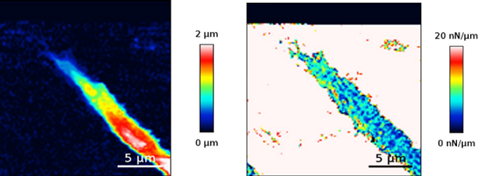
AFM scans of a vital axon.
(left) a topography scan. (Right) measurement of Stiffness.